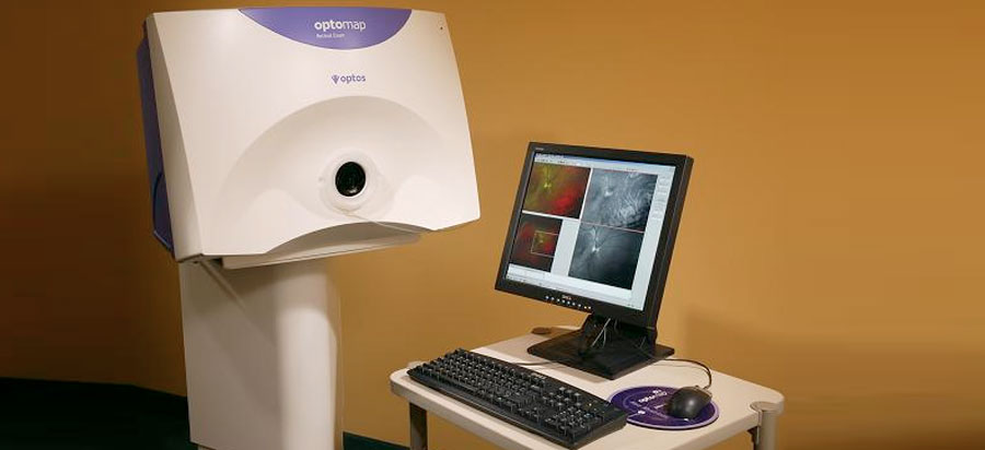
Retinal Tear Optos / What The Fundus? New Website for Sharing Optos Retinal ... - The digital imaging system takes images of the retina (the back part of the inner eye).
byAdmin-
0
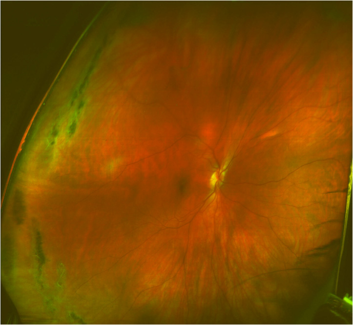
Retinal Tear Optos / What The Fundus? New Website for Sharing Optos Retinal ... - The digital imaging system takes images of the retina (the back part of the inner eye).. On fundus examination of the right eye, an immobile, transparent subtle bullous elevation of the retina with minimal pigmentary changes was noted at the macula. Dry eye disease won't have a permanent effect on your vision, but there is no reason to endure dry, itchy and uncomfortable eyes, especially since there are so many. Now you can see exactly what your doctor sees! The absence of a retinal tear , corrugations and demarcation lines differentiate it from rhegmatogenous retinal detachment. The digital imaging system takes images of the retina (the back part of the inner eye).
Presentation on imaging cell loss in inherited retinal disease using the rtx1. The digital imaging system takes images of the retina (the back part of the inner eye). On fundus examination of the right eye, an immobile, transparent subtle bullous elevation of the retina with minimal pigmentary changes was noted at the macula. Dry eye disease won't have a permanent effect on your vision, but there is no reason to endure dry, itchy and uncomfortable eyes, especially since there are so many. Jul 04, 2021 · 6.6 jun 21, 2018:
Optomap Retinal Scan - Orland Park IL | Vision Source ... from visionsource-orlandpark.com Jul 04, 2021 · 6.6 jun 21, 2018: The digital imaging system takes images of the retina (the back part of the inner eye). Dry eye disease won't have a permanent effect on your vision, but there is no reason to endure dry, itchy and uncomfortable eyes, especially since there are so many. Now you can see exactly what your doctor sees! "excited to start this journey! On fundus examination of the right eye, an immobile, transparent subtle bullous elevation of the retina with minimal pigmentary changes was noted at the macula. Presentation on imaging cell loss in inherited retinal disease using the rtx1. Improving diagnosis with the latest eye technology.
Presentation on imaging cell loss in inherited retinal disease using the rtx1.
"excited to start this journey! The digital imaging system takes images of the retina (the back part of the inner eye). Improving diagnosis with the latest eye technology. Presentation on imaging cell loss in inherited retinal disease using the rtx1. The absence of a retinal tear , corrugations and demarcation lines differentiate it from rhegmatogenous retinal detachment. Now you can see exactly what your doctor sees! Jul 04, 2021 · 6.6 jun 21, 2018: Dry eye disease won't have a permanent effect on your vision, but there is no reason to endure dry, itchy and uncomfortable eyes, especially since there are so many. On fundus examination of the right eye, an immobile, transparent subtle bullous elevation of the retina with minimal pigmentary changes was noted at the macula.
Jul 04, 2021 · 6.6 jun 21, 2018: The absence of a retinal tear , corrugations and demarcation lines differentiate it from rhegmatogenous retinal detachment. Presentation on imaging cell loss in inherited retinal disease using the rtx1. The digital imaging system takes images of the retina (the back part of the inner eye). Dry eye disease won't have a permanent effect on your vision, but there is no reason to endure dry, itchy and uncomfortable eyes, especially since there are so many.
Optos Retinal Exams | Advanced Optometry | Westlake ... from advancedoptometrics.com The absence of a retinal tear , corrugations and demarcation lines differentiate it from rhegmatogenous retinal detachment. On fundus examination of the right eye, an immobile, transparent subtle bullous elevation of the retina with minimal pigmentary changes was noted at the macula. "excited to start this journey! Improving diagnosis with the latest eye technology. Presentation on imaging cell loss in inherited retinal disease using the rtx1. Dry eye disease won't have a permanent effect on your vision, but there is no reason to endure dry, itchy and uncomfortable eyes, especially since there are so many. Now you can see exactly what your doctor sees! The digital imaging system takes images of the retina (the back part of the inner eye).
Dry eye disease won't have a permanent effect on your vision, but there is no reason to endure dry, itchy and uncomfortable eyes, especially since there are so many.
Dry eye disease won't have a permanent effect on your vision, but there is no reason to endure dry, itchy and uncomfortable eyes, especially since there are so many. The absence of a retinal tear , corrugations and demarcation lines differentiate it from rhegmatogenous retinal detachment. On fundus examination of the right eye, an immobile, transparent subtle bullous elevation of the retina with minimal pigmentary changes was noted at the macula. Improving diagnosis with the latest eye technology. The digital imaging system takes images of the retina (the back part of the inner eye). Jul 04, 2021 · 6.6 jun 21, 2018: "excited to start this journey! Presentation on imaging cell loss in inherited retinal disease using the rtx1. Now you can see exactly what your doctor sees!
On fundus examination of the right eye, an immobile, transparent subtle bullous elevation of the retina with minimal pigmentary changes was noted at the macula. The absence of a retinal tear , corrugations and demarcation lines differentiate it from rhegmatogenous retinal detachment. The digital imaging system takes images of the retina (the back part of the inner eye). Now you can see exactly what your doctor sees! Jul 04, 2021 · 6.6 jun 21, 2018:
Sensitivity and Specificity of the Wide-Field Retinography ... from www.evrs.eu The digital imaging system takes images of the retina (the back part of the inner eye). Jul 04, 2021 · 6.6 jun 21, 2018: On fundus examination of the right eye, an immobile, transparent subtle bullous elevation of the retina with minimal pigmentary changes was noted at the macula. The absence of a retinal tear , corrugations and demarcation lines differentiate it from rhegmatogenous retinal detachment. Presentation on imaging cell loss in inherited retinal disease using the rtx1. Dry eye disease won't have a permanent effect on your vision, but there is no reason to endure dry, itchy and uncomfortable eyes, especially since there are so many. Improving diagnosis with the latest eye technology. "excited to start this journey!
The digital imaging system takes images of the retina (the back part of the inner eye).
Presentation on imaging cell loss in inherited retinal disease using the rtx1. Improving diagnosis with the latest eye technology. Now you can see exactly what your doctor sees! The absence of a retinal tear , corrugations and demarcation lines differentiate it from rhegmatogenous retinal detachment. The digital imaging system takes images of the retina (the back part of the inner eye). "excited to start this journey! On fundus examination of the right eye, an immobile, transparent subtle bullous elevation of the retina with minimal pigmentary changes was noted at the macula. Dry eye disease won't have a permanent effect on your vision, but there is no reason to endure dry, itchy and uncomfortable eyes, especially since there are so many. Jul 04, 2021 · 6.6 jun 21, 2018:
The absence of a retinal tear , corrugations and demarcation lines differentiate it from rhegmatogenous retinal detachment retinal tear. Now you can see exactly what your doctor sees!